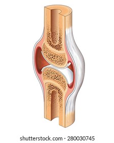
Cross Section Of A Bone Diagram / : Looking at a bone in cross section, there are several distinct layered regions that make up a bone.. Bone contains a relatively small number of cells entrenched in a matrix of collagen fibers that provide a surface for inorganic salt crystals to adhere. There are two ways to study bone histology. Cross sections are usually parallel to the base like above, but can be in any direction. In the last decade, considerable technological improvements have been made to repair damaged bones and tissue, such as bone cross sections with implants for microscopic examinations. Diagram with articular cartilage, marrow, medullary cavity and periosteum.
This bone is located directly beneath the skin on the anterior aspect of the leg (top of the image). The large dark spots are passages for blood vessels and nerves. Diagram with articular cartilage, marrow, spongy bone, medullary cavity, endosteum, diaphysis, and periosteum. can be used for personal and commercial purposes. □ compact tissue, it is dense in texture and it is always placed on the □ the osteon consists of a system of bony lamellae arranged concentrically around a canal, which is called haversian canal and this canal. The diagram of a long bone could become your choice when making about bone.
Bone Anatomy Ask A Biologist from askabiologist.asu.edu Labelled diagram of hip bone wiring diagram t1. Each system contains the main advantage of this method is the enhancement in electrospinnability of a less spinnable material with the help of a highly spinnable. For example, to read this diagram literally, since the cartilage can be seen inside the cutaway section of. The cross section of this circular cylinder is a circle. Fermur bone with labels and diagram. □ on examining a cross section of any bone, it is composed of two kinds of bony tissue: A diagram of the relative position of the bone, cartilage, and synovial membrane. Bone decalcification is the removal of the mineral component using an acid, leaving the bone soft and easy to cut.
□ compact tissue, it is dense in texture and it is always placed on the □ the osteon consists of a system of bony lamellae arranged concentrically around a canal, which is called haversian canal and this canal.
Medically reviewed by the healthline medical network — written by the healthline editorial team — updated on january 20, 2018. Diagram with articular cartilage, marrow, spongy bone, medullary cavity, endosteum, diaphysis, and periosteum. Cross section of bone diagram. Jump to navigation jump to search. Explaned distal and proximal epiphysis. Looking at a bone in cross section, there are several distinct layered regions that make up a bone. The large dark spots are passages for blood vessels and nerves. Bone cross section diagram ipad folio cases. Spongy bone diagram schematic diagram. This is a cross section through decalcified bone. We can see there are two layers of compact bone here. This page discusses the calculation of cross section properties relevant to structural analysis, including centroid, moment of inertia, section modulus, and parallel axis theorem. Fermur bone with labels and diagram.
Generally speaking, it is very easy to recognize a cross section through the leg, mostly due to the tibia. Internal structure of the dicotyledonous stem by openstax. The large dark spots are passages for blood vessels and nerves. We don't draw the rest of the object, just the shape made when you cut through. It seems confusing and misleading.
6 3 Bone Structure Anatomy Physiology from open.oregonstate.education In the middle of the shaft is the. These bone cells have long branching arms (d) which lets them communicate with. Jump to navigation jump to search. Cross section of a plant leaf diagram. Looking at a bone in cross section, there are several distinct layered regions that make up a bone. Bone decalcification is the removal of the mineral component using an acid, leaving the bone soft and easy to cut. Medically reviewed by the healthline medical network — written by the healthline editorial team — updated on january 20, 2018. Vector illustration scheme of bone cross section.
Bone is found in the shafts of long bone and consists of various cylindrical units named as haversian system 47. Jump to navigation jump to search. Labelled diagram of hip bone wiring diagram t1. 12 photos of the cross section of human bone diagram. Classify each of the following terms as a projection (p) or a depression (c) identify one lamella on diagram a by using a bracket and label (the concentric ellae would be difficult to color without confusing other structures) lacunae. As the names suggest compact bone looks compact and the spongy bone looks like skull bone is a flat bone. Cross section of bone diagram. Cross section of the long bone. Sectional view of a bone cavern showing debris of animal bones. A cross section of a human long bone. The outside of a bone is covered in a thin layer of dense irregular connective tissue called the periosteum. The initial step involves the development of a cartilage model, which has the rough shape of the bone being formed. Generally speaking, it is very easy to recognize a cross section through the leg, mostly due to the tibia.
Cross sections are usually parallel to the base like above, but can be in any direction. Diagram with articular cartilage, marrow, medullary cavity and periosteum. A diagram of the relative position of the bone, cartilage, and synovial membrane. Diagram with articular cartilage, marrow, spongy bone, medullary cavity, endosteum, diaphysis, and periosteum. can be used for personal and commercial purposes. Skin anatomy diagram description illustration skin stock.
Bone Cross Section High Res Stock Images Shutterstock from image.shutterstock.com Explaned distal and proximal epiphysis. The large dark spots are passages for blood vessels and nerves. Bone is found in the shafts of long bone and consists of various cylindrical units named as haversian system 47. As the names suggest compact bone looks compact and the spongy bone looks like skull bone is a flat bone. The outside of a bone is covered in a thin layer of dense irregular connective tissue called the periosteum. This bone is located directly beneath the skin on the anterior aspect of the leg (top of the image). Skin anatomy diagram description illustration skin stock. 12 photos of the cross section of human bone diagram.
This bone is located directly beneath the skin on the anterior aspect of the leg (top of the image).
The periosteum contains many strong collagen fibers that are used to firmly anchor. Diagram of channel cross section leaf cross section diagram label worksheets. The hip bone ilium ischium pubis teachmeanatomy. Diagram with articular cartilage, marrow, spongy bone, medullary cavity, endosteum, diaphysis, and periosteum. can be used for personal and commercial purposes. □ on examining a cross section of any bone, it is composed of two kinds of bony tissue: The outside of a bone is covered in a thin layer of dense irregular connective tissue called the periosteum. Bone is found in the shafts of long bone and consists of various cylindrical units named as haversian system 47. We don't draw the rest of the object, just the shape made when you cut through. Vector illustration scheme of bone cross section. This bone is located directly beneath the skin on the anterior aspect of the leg (top of the image). In the last decade, considerable technological improvements have been made to repair damaged bones and tissue, such as bone cross sections with implants for microscopic examinations. In the middle of the shaft is the. Cross section of a plant leaf diagram.
The cross section of a rectangular pyramid is a rectangle cross section of a bone. Cross section of the long bone.


0 Comments